There are many different types of glaucoma. However, most glaucomas can be divided into two categories:
- open-angle glaucoma; where the drain of the eye is wide open, and
- closed-angle glaucoma; where the drain of the eye is partially or fully obstructed.
Within these categories there are different variants of open angle and angle-closure glaucoma which can be classified according to cause:
- Primary glaucomas: No known cause, occurs in susceptible individuals.
- Secondary glaucomas: Another disorder or problem within the eye (such as injury, surgery, drugs or other ocular diseases) causes the glaucoma.
Clinicians may also refer to someone as a glaucoma suspect if they think the person might be showing early signs of glaucoma but they are not yet sure. Many people suspected of having glaucoma at this stage turn out not to have it at all, but some do develop it in time and it is these people who can benefit the most from timely treatment.
The two most common types of glaucoma are Primary Open Angle Glaucoma and Primary Angle-Closure Glaucoma.
Primary Open Angle Glaucoma (POAG)
What is it?
Primary open angle glaucoma (POAG) is the most common form of glaucoma in Australia. It is a condition in which the optic nerve is damaged leading to loss of peripheral vision. There is no cure, however there are treatment options to prevent further loss.
Causes
The damage is usually caused by high intraocular pressure (IOP) within the eye. The rise in pressure is usually due to impaired drainage of fluid out of the eye. The increased pressure damages the optic nerve.
Who is at risk?
Individuals over 40 years with a family history of glaucoma.
Symptoms
Most patients with primary open angle glaucoma have no symptoms of the condition. There is no pain and vision seems normal.
Detection
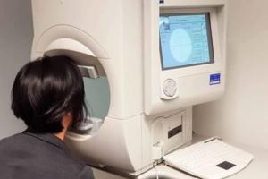
Your eye specialist will conduct a thorough ocular assessment to diagnose POAG. This will include examination of the nerve where it leaves the eye (the optic disc), a check of the intraocular pressure and a field of vision test. Other tests may include measurement of the central corneal thickness and an examination of the drainage angle with a special lens placed on the eye.
Treatment
Damage caused by glaucoma cannot be reversed but treatment can control the condition. If POAG is not treated then people with glaucoma will slowly lose their peripheral vision. Over time, central vision may decrease and eventually this can lead to blindness.
POAG is usually treated with eye drops that reduce the intraocular pressure. It is important to take your eye drops as instructed by your eye care professional. If the eye drops cannot control the intraocular pressure to a sufficient level to stop the loss of visual field your doctor may recommend laser or surgery.
Ongoing Management
As with other types of glaucoma, regular review by an eye specialist is critical to ensure that you do not develop substantial vision impairment.
Additional Information
Glaucoma is an inherited condition so be sure to tell your first-degree relatives that you have the condition so they can have their eyes reviewed.
Acute Angle-Closure Glaucoma
What is it?
Glaucoma of this type is the second most common and involves a narrow drainage angle. In this case, the iris (coloured part of the eye) is usually too close to the drainage angle and can block the passage for the fluid to pass through. If angle closure occurs suddenly, the IOP may rise abruptly causing an ACUTE angle-closure glaucoma attack. Acute angle-closure is a medical emergency requiring urgent treatment.
Causes
This type of glaucoma is the result of an inherited narrowness of the drainage angle of the eye. The angle is located between the iris (the coloured part of the eye) and the trabecular meshwork through which aqueous fluid must drain to leave the eye. As the lens of the eye grows throughout life, the tendency to drain-narrowing becomes more marked with advancing years. When the pupil dilates (with dim lighting conditions, with strong emotions, or with the use of certain types of medications), the contact between the iris and the lens resists the forward flow of aqueous, pressure builds up behind the iris forcing it onto the trabecular drain. This blocks the flow of the aqueous out of the eyes; the pressure rises rapidly. It may reach 60 or 70 mm Hg (the same units used to measure blood pressure) - instead of the usual level of 10 to 20.
Who is at risk?
It is more common in long-sighted eyes, older people, women and in Asian populations.
Symptoms
This sudden rise in pressure produces dramatic symptoms: blurred vision, coloured rings around lights, severe pain in and around the eye, redness, nausea and possibly vomiting. Although both eyes may be at risk of developing this acute attack (because the shape of the two eyes is usually similar and it is the shape that determines the risk), it is very rare for both eyes to be affected at the same time.
50% of patients with an acute angle closure attack give history of previous intermittent attacks, e.g. episodes of blurring of vision lasting 1- 2 hours, associated with haloes around lights, eye ache or frontal headache.
Detection
The symptoms of acute angle closure are severe and usually compel a person to seek medical help. An eye care provider will make the diagnosis of the disease.
Treatment
To preserve vision, the high pressure must be reduced quickly. Drops like pilocarpine, timolol, brimonidine as well as tablets and intravenous medications accomplish this. A peripheral iridotomy is performed with a laser. A laser iridoplasty may need to be performed if a peripheral iridotomy cannot be done. Usually a similar iridotomy is performed in the other eye too, to protect it from developing a similar attack.
Ongoing Management
Once the iridotomy has been performed, the eye pressure is often normal, and medications are not needed in the longer term. However, if the drain has been damaged by the acute attack, it may not be able to work efficiently again. If this is the case, drops may then be required. The eyes must be checked regularly and indefinitely.
Primary Angle-Closure (PAC) and Primary Angle-Closure Glaucoma (PACG)
What is it?
Primary angle-closure (PAC) is defined as appositional or synechial closure of the anterior chamber angle which can lead to aqueous outflow obstruction and raised eye pressure, in the absence of glaucomatous optic neuropathy. PAC is generally bilateral. Optic nerve damage resulting from PAC is described as primary angle-closure glaucoma (PACG).
Causes
Primary angle-closure is the result of an inherited narrowness of the drainage angle of the eye. The angle is located between the iris (the coloured part of the eye) and the trabecular meshwork through which aqueous fluid must drain to leave the eye. As the lens of the eye grows throughout life, the tendency to drain-narrowing becomes more marked with advancing years.
Primary angle-closure may develop after an acute episode of angle-closure with persistent adhesions that block the trabecular meshwork and aqueous drainage (peripheral anterior synechiae). More commonly it develops following gradual asymptomatic closure of the angle.
Who is at risk?
It is more common in long-sighted eyes, older people, women, and in Asian populations.
Symptoms
Typically PAC and PACG develop chronically without symptoms, however an acute rise in IOP Acute angle closure (AAC) crisis (unilateral in 90% of cases) can present as a clinical emergency (see separate information segment titled Acute Angle-Closure).
Detection
An optometrist will often make the diagnosis based on a routine examination in a patient without symptoms.
Treatment
Intraocular pressure control in patients with primary angle-closure depends on the amount of trabecular meshwork damage and extent of angle closure.
A peripheral iridotomy is performed with a laser in eyes with narrow angles to prevent further closure and blockage of the trabecular meshwork. A laser iridoplasty may need to be performed if the angle remains narrow. Lens extraction can be utilised to eliminate the narrow angle as well.
If the drain has been damaged or is blocked, it may not be able to work efficiently. If this is the case, medical treatment in the form of drops or surgery may be required. Drops are used to reduce the intraocular pressure. Trabeculectomy surgery may rarely be needed in cases where the extent of angle closure is extensive and intraocular pressure control is difficult even with drops.
Ongoing Management
The eyes must be checked regularly and indefinitely in all cases of primary angle-closure and primary angle-closure glaucoma.
Additional Information
Secondary angle-closure as opposed to primary angle-closure is used to describe raised eye pressure due to narrow angles caused by certain types of medication and following eye surgery.
Normal Tension Glaucoma
What is it?
The term glaucoma refers to a characteristic pattern of damage to the optic nerve (the nerve that enters the back of the eye and carries the visual signal to the brain) along with corresponding visual field loss which is progressive. Various risk factors for the development of glaucoma are recognised, but many other risk factors remain unknown.
One of the main risk factors for the development of glaucoma is the intraocular pressure (the pressure within the eyeball). The higher the pressure, the more likely glaucoma is to develop. The normal eye pressure is 10-21 mmHg - eye pressure is measured on the same scale as blood pressure. However this is not the only risk factor for glaucoma. It is also widely recognised that in about 1/3rd of cases of glaucoma the characteristic optic nerve changes and visual field loss can develop in an eye with normal pressure – this is termed normal tension glaucoma.
Causes
A history of migraine and Raynaud’s phenomenon is linked to normal tension glaucoma. It is thought that lower blood pressure or reduced ocular blood flow may render the optic nerve more susceptible to the development or progression of normal tension glaucoma.
Who is at risk?
Individuals over 40 years with a family history of glaucoma and a history of migraine or Raynaud’s phenomenon.
Symptoms
Most patients with normal tension glaucoma in the early stages have no symptoms of the condition. There is no pain and vision seems normal.
Detection
The diagnosis of glaucoma requires recognition of the characteristic optic nerve and visual field changes. This requires examination of the back of the eye and testing of the visual field. There are also tests that can help measure the optic nerve and nerve fibres as they enter the back of the eye.
Patients with normal tension glaucoma form a most important group, their evaluation needs to be especially thorough both to confirm the diagnosis and to exclude other medical conditions that may mimic glaucomatous damage to the optic nerves. They are of research interest because they demonstrate that factors other than an abnormally raised eye pressure can cause glaucomatous nerve damage - there is much work underway around the world to identify these factors so they can be treated.
Treatment
Even though the pressure is still in the normal range, there is good evidence to show that lowering the pressure with eye drops (and sometimes laser or surgery) can help slow the process and preserve patients’ vision. The range of treatments is thus similar to those for the more common forms of glaucoma, but often aiming for lower pressures. As with all forms of glaucoma, early detection and prompt, effective treatment is vital to reduce visual damage.
Ongoing Management
As with other types of glaucoma, regular review by an eye specialist is critical to ensure that you do not develop substantial vision impairment.
Childhood Glaucoma
What is it?
Childhood Glaucoma describes the different types of glaucoma sometimes referred to separately as Congenital, Infantile or Juvenile glaucoma. There is usually an abnormal development of the eye's aqueous fluid outflow system. Most patients with this condition come under treatment in infancy or early childhood, however, some may not have their glaucoma diagnosed until adulthood.
Childhood glaucoma is uncommon but when it occurs the effect on vision can be devastating. Early diagnosis and appropriate treatment can make a significant difference to the child's sight. There is a large variation in severity in this subtype of glaucoma, some mild and others severe, however any vision during a child's development is worth fighting for.
Causes
There are three major types of childhood glaucoma:
- Developmental diseases of the eye, where the aqueous fluid drainage pathways are imperfectly developed;
- Eye diseases such as inflammation, or an injury that can lead to glaucoma and;
- Glaucoma that follows cataract surgery as a young child (usually <1y.o.)
Who is at risk?
There are several genes and inheritance patterns in childhood glaucoma. In general and in the absence of a family history, the chance of two unrelated parents with an affected child having another affected baby is between 3% and 15%. If other family members have glaucoma or parents are related the risk may be much higher. If a child is diagnosed as having glaucoma, then all brothers and sisters need to be examined as well. A consultation with a clinical geneticist and possibly genetic testing can be arranged to further detail these risks to other family members.
Symptoms
How does childhood glaucoma reveal itself? High eye pressure in babies stretches their softer eyeball so that the eye increases in size. The child often dislikes light and, if older, may bury its head into a parent’s arms to escape light. Watering of the eye is also often noticed. Sometimes the condition is only noticed when the deepest layer of the stretched cornea splits, with the aqueous fluid entering the cornea, turning it cloudy or white. If the glaucoma is particularly severe the child may be born with cloudy corneas, indicating that the eye pressure has been significantly raised in the womb.
Detection
Assessment of the possibly-affected child can be difficult in babies or little children who usually do not co-operate, thus examination under anaesthetic is often required. In examination, the size and shape of the eye is measured, the eye pressure is recorded and the structures of the tissues in the front and back of the eye are
assessed.
Once a firm diagnosis has been made and the baseline measurements are recorded, treatment can be planned. Inflammation, injuries and growths that are the cause of the glaucoma are treated specifically and these measures should lower the eye pressure.
Treatment
If there is an abnormal development of the drainage pathways this may be managed firstly by drops and/or liquids by mouth to reduce eye pressure, and then by surgery. If the cornea (the window of the eye) is normal in structure and clear, a goniotomy is usually performed.
This consists of passing a special knife-needle across the front of the eye (the anterior chamber) and gently cutting the abnormal tissue that is blocking the flow of aqueous fluid from draining from the anterior chamber of the eye through the trabecular meshwork (the drainage tissues).
The drain is a ring that extends for 360° around the inside of the front of the eye. In a single goniotomy operation about 120 degrees of the abnormal tissue can be cut. If one operation does not lower the eye pressure sufficiently, a second and even a third goniotomy may be needed for each eye.
If the cornea is cloudy and the surgeon cannot see across the anterior chamber to cut the abnormal tissue, then another operation called a trabeculotomy is needed. In this procedure the tiny canal into which the aqueous should be flowing is identified, an instrument is inserted into it and delicately swept into the anterior chamber. This opens the drain into the anterior chamber, and allows the aqueous fluid an easier exit. A trabeculotomy too may need to be performed more than once to lower eye pressure effectively.
Occasionally these operations cannot be performed for technical reasons, or they do not succeed in reducing the eye pressure adequately. Then either a trabeculectomy drainage operation or the placement of an artificial plastic tube and plate on the outside of the eye is needed. Each operation depends on the condition of each eye.
Ongoing Management
The success of any of these operations has to be reviewed for the life of the child. Initially this means regular checks and sometimes with examinations under anaesthesia until the child can co-operate fully. On each occasion the structures of the tissues of the eye and the pressure are measured. The cornea should be clear and it should only be increasing in size in line with expected growth. The eye pressure should be in the normal range of 10-20 mm Hg, and the shape of the nerve fibres in the optic disc at the back of the eye should be stable.
The need for glasses is assessed and if one eye is affected more severely than the other, the child may need to wear an eye patch for a period of time each day. The end result sought is not just a normal eye pressure (although this is the first important goal without which nothing much else can be achieved) but a well-seeing eye in a happy child. Treatment of childhood glaucoma is therefore complex and care and attention is needed for the life of the patient. There are no short-cuts. The range of severity of these diseases is wide. Some affected children will achieve and maintain normal vision; some will achieve partial but effective sight, whereas some will lose their sight no matter what is tried for them.
Pigment-Dispersion Syndrome
What is it?
Glaucoma is usually caused by an increase in intraocular pressure (IOP) which can damage the optic nerve. Pressure can be elevated if the trabecular drainage tissues become blocked by particles of pigment.
Causes
The focusing lens of the eye is held in position by scaffolding called the zonule. If the iris (the coloured part of the eye) bows backwards, it can rub against these zonules. The pigment granules, which give the iris its colour, may be dislodged. They flow with the aqueous fluid until the trabecular mesh traps them as the fluid leaves the eye – much like a strainer traps tea leaves. As more and more pigment granules become caught, the trabecular drain works less and less efficiently. The eye pressure starts to rise. Some eyes tolerate a great deal of pigment dispersion before this occurs; other eyes are not so fortunate.
Who is at risk?
Eyes that are modestly short-sighted are the ones most commonly affected by pigment dispersion. Perhaps this is because their shape lends itself to rubbing between iris and zonule. We do not know why men are affected more than women, or why it classically begins before the age of 35 years. Pressure elevation may not follow until many years later but, because it may do so suddenly, patients whose eyes demonstrate pigment dispersion need to be examined regularly.
Symptoms
Eyes with this syndrome can have a sudden release of pigment after pupil dilation or with jarring (e.g. running on hard surfaces). There may be a sudden pressure rise with blurred vision and coloured rings around lights.
Treatment
Your ophthalmologist may perform a procedure using a laser to alter the iris profile in an attempt to avoid the rubbing between the iris and zonule structures. Treatment will be started once the pressure rises. In the first instance this consists of drops. If medical treatment fails to control eye pressure adequately, other laser treatment may also be used. If all this fails to control the pressure at levels safe for the optic nerve, then surgery may become necessary.
Ongoing Management
As with other types of glaucoma, regular review by an eye specialist is critical to ensure that you do not develop substantial vision impairment.
Pseudo-Exfoliation Syndrome and Glaucoma
What is it?
In some eyes with raised intraocular pressure (IOP) and glaucoma, many tiny white flakes can be seen when viewed through a slit lamp microscope, lying on the edge of the pupil, and on the front surface of the crystalline lens. These white flakes have the appearance of microscopic dandruff and are usually accompanied by a mild dispersion of pigment granules from the back surface of the iris, with accumulation of this pigment in the tissues of the trabecular meshwork.
This is different from a condition called pigment dispersion syndrome. About 50% of the time, only one eye of a patient is affected by the pseudo-exfoliation syndrome.
Causes
The deposit of white flakes and pigment granules on the trabecular meshwork interferes with the drainage of aqueous fluid from the inside of the eye to the blood vessels on the surface of the globe. The eye pressure then rises. It may do so rapidly and to very high levels. If this occurs damage to the optic nerve fibres and thus to the vision, may occur rapidly.
Who is at risk?
The condition is more prevalent in Northern Europeans particularly those of Scandinavian descent and in individuals aged greater than 60years.
Symptoms
Generally glaucoma in the early stages has no symptoms. However occasionally an individual may notice an eye ache and/or frontal headache and/or blurry vision. As the glaucoma becomes more advanced patches of persistent blurry vision may be observed.
Detection
Any eye with pseudo-exfoliation syndrome is at risk of developing glaucoma, even if the pressure has not risen at the time the condition is first detected. All eyes with pseudo-exfoliation, even with normal pressure, need to be checked by an ophthalmologist regularly. The patient usually does not feel a rise in pressure, and it is only by pressure testing that it can be detected.
Treatment
If the eye pressure does rise, treatment aims to lower the pressure to prevent the onset of glaucoma or prevent it getting worse. In the first instance, this is usually with either medical treatment or a procedure called selective laser trabeculoplasty. With medical treatment, one or more types of drops are used to slow down the pumping of aqueous into the eye, or to speed up its drainage. Often, however, pseudo-exfoliation glaucoma is resistant to medical therapy. Fortunately, this condition responds to SLT about 85-90% of the time, compared with a 70-75% response rate of "ordinary" glaucoma to this type of laser. In some eyes, the pressure-lowering effect of the laser (with or with-out eye drops) can last for many years.
If SLT and/or medical treatment fail to control eye pressure, then drainage surgery, similar to that for other types of glaucoma, can be employed. The success rate for surgery is the same as for other forms of glaucoma - about 80-85%, unless there are special complications applicable to a particular eye.
Ongoing Management
As with other types of glaucoma, regular review by an eye specialist is critical to ensure that you do not develop substantial vision impairment. Other important information to consider; This condition runs in families. If you have a relative with this condition and you are forty years old or older, a review by an eye care provider is recommended.
Neovascular (Rubeotic) Glaucoma
What is it?
The normal eye has a very high demand for nutrients such as oxygen to allow it to function normally. Various processes ensure the delivery of oxygen to these tissues via the ocular circulation. However some diseases may result in the eye being starved of blood and oxygen which may lead to a cascade of processes finally resulting in neovascular glaucoma.
Causes
In Australia the most common causes of this are retinal vein occlusion, advanced diabetic eye disease and ocular ischaemic syndrome. In retinal vein occlusion there is a blockage in the main venous drainage from the retina, resulting in very poor circulation. Severe narrowing of retinal arterioles in advanced diabetic retinopathy may also lead to poor delivery of blood and oxygen to the retina.
Ocular ischaemic syndrome describes a condition where there is poor delivery of blood to the eye as a whole, usually due to severe narrowing of the carotid artery in the neck. When this occurs, cells within the retina become stressed and, in an attempt to overcome the lack of oxygen, they produce a range of chemicals known as angiogenic factors. These chemicals diffuse through the retina and into the front of the eye with their main effect to cause new blood vessels to grow. Elsewhere in the body this may help to overcome the ischaemia but in the eye it has disastrous results.
Who is at risk?
Individuals 40 years and older with diabetes, high cholesterol and high blood pressure who have experienced a retinal vein occlusion or have diabetic retinopathy.
Symptoms and Detection
Under this influence new vessels begin to grow on the surface of the iris, with a fibrotic membrane eventually growing across the drainage channels in the front of the eye, blocking them completely. This results in a sudden and extreme rise in intraocular pressure, making the eye very painful. The conditions underlying this neovascular process by themselves are often associated with poor vision but the additional insult of very high intraocular pressure may damage what residual vision there is.
Prompt treatment is necessary to try to salvage any remaining vision as well as to make the eye more ‘comfortable’.
Treatment
Treatment is two-fold: firstly controlling the intraocular pressure and secondly identifying the cause of and treating the neovascular process. These treatments are undertaken concurrently.
Initially, medical treatment with eye drops to decrease aqueous fluid production is commenced to lower intraocular pressure. Often additional oral agents such as acetazolamide are needed, because the pressure can be very difficult to control. Even with multiple medications, adequate pressure control may not be achieved and frequently, laser or surgery may be required.
Controlling the neovascular process involves trying to switch off the production of the stress response chemicals or angiogenic factors. Significant advancements in the understanding of these agents have occurred over the past, resulting in the development of drugs such as Bevacizumab. This is a manufactured antibody that neutralises the angiogenic factor. Given into the eye by injection it can rapidly turnoff the neovascular process but is not a complete treatment in itself.
To stop further production of the angiogenic factors, areas of ischaemic retina need to be treated with laser; or if the problem is due to carotid artery disease this needs to be addressed by vascular surgery.
If medical treatment mentioned above is not sufficient to control the intraocular pressure, as is not uncommon, surgery or cyclodiode laser may be required. The approach taken is dependent on the level of remaining vision. If there is reasonable salvageable vision surgery such as trabeculectomy or a tube/shunt procedure may be advised. These both create an artificial channel to allow aqueous fluid to bypass the blocked drainage channels. In the tube procedure aqueous drains out of the eye via a fine silicone tube to a reservoir created by a plate secured to the white of the eye.
If the potential for vision is poor then cyclodiode laser may be performed with a series of treatments placed over the region of the ciliary body at the front of the eye. This area is responsible for the production of the aqueous fluid and by selectively destroying some of this tissue with the laser the amount of aqueous fluid produced decreases, allowing the intraocular pressure to decrease.
In summary, this is an aggressive type of glaucoma which often has a poor outcome, as vision is frequently damaged by the underlying cause of the neovascularisation. The main goals of treatment are to preserve the remaining sight and to leave the patient with a comfortable eye.
Ongoing Management
As with other types of glaucoma, regular review by an eye specialist is critical to protect against further visual loss
Mixed Mechanism Glaucomas
What is it?
Combined mechanism glaucomas refers to an eye with elevated pressure in which both open-angle and angle closure glaucoma mechanisms are present. Usually the structure of the eye is similar to that found in eyes with angle closure glaucoma – that is; the globe is somewhat smaller than ideal, with the result that the internal ocular components are crowded together.
This means the coloured part of the eye (the iris) is very close to the trabecular drainage tissues, making it difficult for the aqueous fluid to gain access to the drain, and raising the possibility that the iris tissues can completely block the drain.
Who is at risk?
It is more common in long-sighted eyes, older people, women, and in Asian populations
Symptoms
A sudden rise in pressure produces dramatic symptoms: blurred vision, coloured rings around lights, severe pain in and around the eye, redness, nausea, and possibly vomiting.
Treatment
A peripheral iridotomy procedure, usually performed with a laser, permits the iris to fall back, away from the drain, allowing the aqueous to access the drain and thereby permitting the pressure to fall to normal levels.
However, if iris tissue (because of its previous proximity) has blocked the drain intermittently or repeatedly, the drainage system may be functionally impaired. Consequently, this will abnormally resist the drainage of aqueous even when opened by laser peripheral iridotomy.
In the case of mixed mechanism glaucomas the affected eye also has an open-angle glaucoma mechanism causing raised pressure, so that once the angle closure component has been eliminated, the drain still does not operate efficiently and the pressure remains too high for the safety of the optic nerve fibres.
Long-term drops, laser treatment to the drain itself, or even drainage surgery may become necessary to preserve sight, just as in the open-angle glaucomas.
These situations are known as "combined mechanism glaucomas". Often the diagnosis can only be made once the angle closure element has been removed, and the pressure subsequently monitored. It means that when angle closure has been treated successfully long-term care and observation remain vital to ensure that should open angle glaucoma occur it is promptly detected thus minimising the risk of unnecessary blindness.
Ongoing Management
As with other types of glaucoma, regular review by an eye specialist is critical to ensure that you do not develop substantial vision impairment
Traumatic Glaucoma
What is it?
Traumatic glaucoma refers to cases in which a direct injury to the eye has led to the development of glaucoma.
Glaucoma is the name given to a group of eye diseases in which the optic nerve at the back of the eye is slowly destroyed. The optic nerve carries visual information from the eye to the brain. In most people this damage is due to an increased pressure inside the eye ‐ a result of blockage of the circulation of fluid within the eye, or its drainage.
In other patients the damage may be caused by poor blood supply to the vital optic nerve fibres, a weakness in the structure of the nerve, and/or a problem in the health of the nerve fibres themselves.
Causes
Blunt or penetrating trauma may cause a rise in pressure inside the eye. This raised pressure may go on to cause glaucoma straight away or take some time. A blow to the eye such as a punch, car accident or head injury can cause damage to the drainage mechanism in the eye. This may be seen as bleeding, or inflammation within the eye; or there may be direct damage to the drainage pathway, or to the optic nerve itself.
This generally settles with initial treatment, but some people may develop a late rise in the pressure inside the eye. Unlike the initial injury, this is not often painful as the pressure creeps up over several years and this high pressure is the major treatable cause of the damage to the optic nerve in glaucoma.
Who is at risk?
Six months after a blunt injury 3.4% of people may develop glaucoma. The risk is increased if there was:
- bleeding inside the eye, poor vision, or injury to the lens
- damage to the fluid drainage pathway of the eye.
Older patients are also at increased risk. (Sihota, Sood et al. 1995, Girkin, McGwin et al. 2005)
A penetrating injury where something foreign goes through the wall of the eye carries a 2.7% risk of developing glaucoma. (Girkin, McGwin et al. 2005)
If there was damage to the fluid drainage in the eye, glaucoma develops in 9% of people with long‐term follow‐up. (Kaufman and Tolpin 1974)
Symptoms
Generally glaucoma in the early stages has no symptoms. However occasionally an individual may notice an eye ache and/or frontal headache and/or blurry vision.
As the glaucoma becomes more advanced patches of persistent blurry vision may be observed.
Detection
Glaucoma seems to occur in two peak periods, around 3 and 10 years after a traumatic injury (De Moraes and Susanna Jr 2016). For this reason it is important to continue to monitor the pressure and optic nerve in your eyes if you have had a traumatic injury.
Treatment
Treatment is similar to other glaucomas:
- Medical management with topical drops is most common
- Lasers may occasionally be used to help reduce pressure
- Surgery is an effective option for some people to keep their ocular pressure and optic nerve damage under control.
Ongoing Management
The key here is regular follow up ideally annually by an eye care provider after eye trauma to detect and treat glaucoma early should it arise. Remember glaucoma usually has no symptoms in the early stages.
Other important information to consider
Retinal detachment is also a risk after eye trauma and can occur months after the initial trauma. If you develop symptoms such as black spots, flashing lights or a curtain of darkness in you vision then seek an urgent eye review.
Iridocorneal Endothelial Syndrome
What is it?
The cornea is the clear window in front of the coloured part of the eye (the iris). It has five layers to it and the inner layer is called the endothelium. The endothelium is a single layer of cells that does not regenerate. Its purpose is to pump fluid out of the cornea, effectively preventing it from becoming waterlogged.
In iridocorneal endothelial (ICE) syndrome, there is a dysfunction with this inner layer of cells. Instead of being a single layer of non-regenerating cells, it starts to replicate and spread. This has three main effects in the eye:
- It damages the cornea – instead of being a single layer of cells covering the inside of the cornea, the endothelial cells become multilayered. This results in decreased function of this layer and the cornea becomes waterlogged (oedema), decreasing vision. There are other corneal signs an ophthalmologist will detect that will help confirm the diagnosis of ICE syndrome.
- It causes glaucoma – the eye makes and drains fluid (aqueous). The point where the aqueous drains is called “the angle” and it is located at the junction of the cornea and the iris. Because in ICE syndrome the endothelium replicates and spreads, it eventually covers and damages the angle, preventing the aqueous from draining. This causes the pressure in the eye to increase, and long term high pressure in the eye damages the nerve that connects the eye to the brain (the optic nerve). This is glaucoma and glaucoma damage is irreversible.
- It damages the iris – As the abnormal endothelium replicates and spreads, it eventually starts to grow over the iris. This damages the iris, and can cause the pupil (the black central part of the eye) to stop working in the light, to have an oval shape, or even for the iris to develop multiple defects or holes in it. This can cause glare in the eye.
Causes and who is at risk
No one knows for sure what causes ICE syndrome. It is sporadic in presentation and not passed on genetically (so it cannot be passed on from parent to child). It is more common in women and is most frequently detected between the ages of 20 and 50 years. It almost always only affects one eye.
Symptoms
The initial presentation of ICE syndrome patients may be due to monocular pain (from corneal edema or elevated intraocular pressure from angle-closure), blurry vision, or iris changes.
Detection
Your eye specialist will conduct a thorough ocular assessment to diagnose the disease.
Treatment
Lowering the pressure with eye drops (and sometimes laser or surgery) can help slow or stop the process of glaucoma and preserve patients’ vision. The range of treatments is thus similar to those for the more common forms of glaucoma.
Ongoing Management
As with other types of glaucoma, regular review by an eye specialist is critical to ensure that you do not develop substantial vision impairment.
Steroid-Induced Glaucoma
What is it?
Treatment with steroids can elevate the pressure in the eye. This is termed Steroid- Induced Ocular Hypertension. If there is persistent eye pressure elevation then damage to the optic nerve of the eye can occur with corresponding damage to the peripheral vision, this is termed Steroid Induced Glaucoma.
Steroid hormones are naturally produced by the body but can also be artificially synthesised as medications. Steroid medications are predominantly used as anti- inflammatory treatments for a variety of medical conditions such as asthma, eczema, allergy, rheumatoid arthritis and uveitis to name a few.
They are made in many forms, including as tablets or liquid, as inhalers for the mouth or nose, as topical medications in the form of eye drops, creams and lotions, and finally as injections into blood, muscle, joints or the eye.
Causes
Typically, eye pressure becomes elevated 2 to 4 weeks after use of steroids, and in most cases returns back to normal after cessation of medication, although in a small proportion of people, the eye pressure will remain persistently elevated.
Who is at risk?
Anyone regardless of age exposed to steroids as discussed above.
Symptoms
Generally glaucoma in the early stages has no symptoms. However occasionally an individual may notice an eye ache and/or frontal headache and/or blurry vision. As the glaucoma becomes more advanced patches of persistent blurry vision may be observed.
Detection
If steroid use is going to exceed two weeks then starting 2-3 weeks after steroid commencement, regular review by an ophthalmologist or optometrist is necessary, to assess eye pressure and check for glaucoma. Steroids can also affect other parts of your body such as your blood sugar level, blood pressure and bones, so remember to get a check-up by your general practitioner as well.
Treatment
If the eye pressure becomes elevated, then stopping steroids can often reduce the pressure to normal in 1 to 4 weeks. If eye pressure does not return to normal or steroids cannot be stopped, then eye drops to lower the pressure can be commenced or laser can be used in certain circumstances. Very rarely, an ophthalmologist may need to perform eye surgery to reduce the pressure, if it cannot be controlled by other means. Ideally if safe to do so, alternatives to steroid therapy should always be sought. It may not be possible to avoid steroids as they can be useful medications in many conditions. If avoidance is not possible, then efforts should be directed towards use of steroids with the lowest possible potency, or dosage over the shortest period of time.
Ongoing Management
As with other types of glaucoma, regular review by an eye specialist is critical to ensure that you do not develop substantial vision impairment.
Other important information to consider
Steroids can elevate the eye pressure in any individual. However children, the elderly, persons with glaucoma, glaucoma suspects and immediate relatives of persons with glaucoma are more likely to have a pressure elevation with steroid use. The type of steroid (how potent it is), dosage and mode of administration can affect the steroid response.
Typically, topical use of steroids in the form of eye drops, creams and lotions are more likely to elevate the eye pressure. Steroid tablets or liquid and steroid injections into the blood or eye can also elevate the eye pressure. It is unclear whether inhalers for the mouth can raise the intraocular pressure, with short-term use suggested to be safer than long-term use. It is recommended that those on long-term inhaled steroids or high dose inhaled steroids should be seeking review by an ophthalmologist or optometrist. A recent Australian study has suggested that steroids in the form of nasal spray doesn’t raise the eye pressure.
Uveitic Glaucoma
What is it?
Not all forms of glaucoma are the same. Some types of glaucoma are caused by other diseases in the eye. Some patients experience a condition where there is inflammation within the eye (uveitis) and as a result of this inflammation glaucoma can develop (uveitic glaucoma). Uveitic glaucoma differs in some ways from other forms of glaucoma.
Causes
Uveitis is the term used to describe a group of conditions where there is inflammation in one or more layers of the vascular tissues within the eye. The most common form of uveitis is where the front part of the eye experiences inflammation, and is also termed ‘iritis’ - because it involves the iris (coloured tissue at the front of the eye). Other structures such as the ciliary body (where the clear aqueous fluid comes into the eye) and the choroid (which lies underneath the retina at the back of the eye) can also be inflamed. Inflammation of the eye has a variety of causes. Sometimes it can be caused by an infection, but more commonly the cause is unknown.
The type of uveitis is named based on where the inflammation occurs. In addition to iritis, if it is in the ciliary body it is called cyclitis, and in the choroid it is called choroiditis.
Patients who experience episodes of iritis, or other forms of uveitis, can develop a rise in intraocular pressure as a result of the inflammation or adhesions which form within the eye following inflammation. When raised pressure in the eye causes damage to the optic nerve at the back of the eye, we call this glaucoma.
Treatment
The raised pressure in the eye is treated with eye drops as with other forms of glaucoma. The inflammation must also be treated; usually with anti-inflammatory eye drops, and sometimes with oral medications - depending on severity and response to treatment. If pressure cannot be controlled with medications and ongoing damage to the optic nerve occurs then glaucoma drainage surgery may be required. Trabeculectomy or Glaucoma Drainage Tube are the typical operations used where surgery is required for uveitic glaucoma.
Glaucoma associated with uveitis can be more severe than the more common forms of glaucoma, and can affect people at a younger age than more typical glaucoma cases. Treatment required is sometimes more intensive than other forms of glaucoma, and there is a higher likelihood of needing surgery to control the pressure. Fortunately effective treatments are available - for both the inflammation and the glaucoma. Your eye care provider can work with you to manage this. It is likely to require ongoing treatment and care, though, and be aware that there is no ‘quick fix’.
Treatment must be individualised for the particular situation in your eye. Your eye care provider will be able to answer any specific questions about your condition and treatment plan.
Glaucoma in Older Children
What is it?
Glaucoma in older children (greater than 2 years old) and adolescents is similar to glaucoma in adults. Glaucoma in children of all ages is rare, unlike adult open angle (chronic) glaucoma, which is relatively common. Glaucoma affects approximately 1 in 2000 children.
Causes
Glaucoma in children older than two years is often secondary to other ocular disease or an underlying general disease that affects the child. Examples associated with other ocular diseases are glaucoma following surgery for congenital cataract (aphakic glaucoma) and glaucoma secondary to iritis (inflammation of the iris which in children that is often associated with juvenile chronic arthritis).
Examples of underlying general diseases of childhood that may be associated with the development of glaucoma are neurofibromatosis type 1 (a disorder of the skin/ eye and nerve tissue) and Sturge-Weber syndrome (a vascular birthmark or port wine stain on the face associated with a risk of glaucoma and epilepsy). Primary open angle glaucoma does occur rarely in older children and adolescents.
Who is at risk?
Primary open angle glaucoma in adolescents may show a familial tendency just as in adult open angle glaucoma. Inherited juvenile open angle glaucoma is well recognized but very rare. This form of glaucoma is generally not detectable till the twenties rather than during later childhood.
Most research into the causes and treatment of glaucoma is directed to understanding adult glaucoma. Fortunately many of these advances can be modified to treat children with glaucoma.
Symptoms
In older children the eye responds to increased intraocular pressure in a manner similar to adults. Unlike younger children there is no increase in the size of the eye and the cornea does not become cloudy. Thus the onset of glaucoma in these children is much more likely to be asymptomatic.
Detection
The child needs to be assessed by an ophthalmologist. Ideally the child should be referred to an ophthalmologist with experience in managing childhood glaucoma. The nature of assessment undertaken varies with the age of the child. In children over 7 years the tests used are very similar to those used for adults, i.e. pressure measurement, visual field assessment and examination of the optic disc after dilatation of the pupil.
Treatment
The treatment for glaucoma in older children is generally medical (eye drops) initially and if these fail, surgery is considered. This is similar to the situation with older adults with glaucoma.
Operations such as trabeculectomy or drainage tubes are used. These procedures aim to create a controlled leak or "fistula" by which the aqueous can bypass the trabecular meshwork and escape to drain via the external blood system of the eye.
As with adults anti-inflammatory and antibiotic drops are used post operatively. When trabeculectomy is performed in children an antimetabolite such as 5-fluorouacil (5-FU for short) is very often used as children heal much more rapidly than adults.
Laser trabeculoplasty is rarely used in the treatment of glaucoma in children of any age. A cyclo destructive procedure, such as diode laser treatment of the ciliary body, is sometimes used in the treatment of aphakic glaucoma in children.
Ongoing Management
As in any person if glaucoma is not appropriately treated there is a risk of progressive visual impairment. Rarely does childhood glaucoma result in severe visual impairment but life-long follow up is needed for all children after a diagnosis of glaucoma is made.
In general there is little need to alter lifestyle. Adolescents often have difficulty accepting the need for long-term medication and regular medical review. Ensuring compliance with regular use of eye drops may be especially difficult.

Clinicians will refer to someone as a ‘glaucoma suspect’ if they think the person might be showing early signs of glaucoma and require ongoing monitoring.

Developmental glaucoma is an umbrella term for a glaucoma due to a rare group of eye disorders characterised by abnormal development of the front part of the eye known as the anterior chamber.

Sturge-Weber Syndrome is a congenital condition which can have ocular, neurological and skin manifestations. Glaucoma in Sturge-Weber Syndrome occurs because of the abnormal blood vessels in and around the eye.
It is thought that nearly one third of people with low vision (of any cause) will develop Charles Bonnet Syndrome. In studies of glaucoma, it was found that nearly 1 in 4 patients experienced this phenomenon, which involves visual hallucinations.

“You do have glaucoma.” The words I had been dreading. Four words and I feel as if my feet have been swept out from under me. Even though I’m sitting down, I feel winded and totally devastated.