Glaucoma is a condition arising from progressive and irreversible damage to the optic nerve. The optic nerve is made up of approximately one million nerve fibres called Retinal Nerve fibres (RNF) which connect the back of the eye to the brain and convey visual information.
By viewing the images of each stage of glaucoma, we can better understand the progressive loss of the retinal nerve fibre layer (RNFL), and the visual changes associated with this disease.
Images used to diagnose and monitor glaucoma are:
- Optic nerve photography
- Visual Field Test
- OCT – Optical Coherence Tomography of the Optic Nerve (including a horizontal and vertical view).
Normal eye (no glaucoma)
A normal optic nerve will show a pink, healthy colour peripherally (around the edges) with a lighter coloured area centrally (in the middle), such as seen in the disc photo here. This central area is termed the “cup” and is typically mildly excavated (a dip). This dip can also be seen the horizontal and vertical RNFL scans. Anatomically, the cup is the point at which the retinal ganglion cell axons leave the eye, carrying visual information back to the brain.
The RNFL thickness map shows areas where the retinal nerve fibre layer (RNFL) is thinner (blue), slightly thicker (yellow) and thickest (red). The image shown illustrates a typical pattern of RNFL thickness in a right eye. This is a healthy eye with no glaucoma damage.
This is a normal visual field with no visual field loss. You will note in the normal image there is a dark area at approximately 15 degrees from centre – this is the anatomical blind spot caused by the presence of the optic nerve and it is very normal. Even a healthy eye with no glaucoma damage has this area of blind spot. Apart from the blind spot, put simply, the darker the grey in a visual field image the brighter a light stimulus has to be in order to be seen at this point.
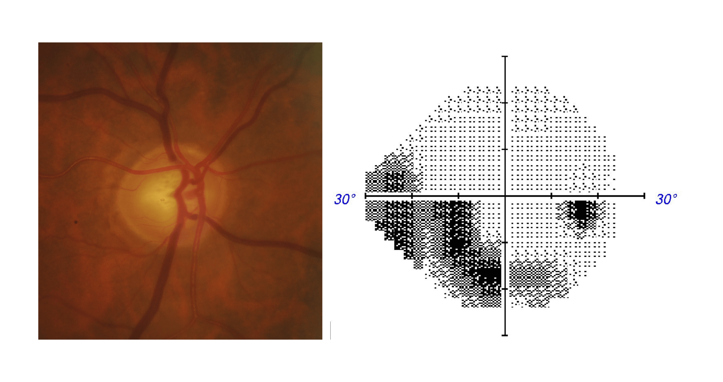
Early glaucoma
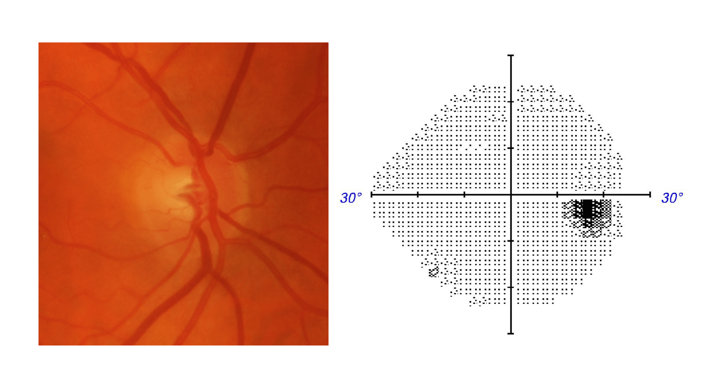
Changes may also be visible at the optic nerve where we typically see a focal expansion of the optic cup, causing thinning (damage to the healthy cells indicated by change in colour) of the pinkish-coloured area around this cup (the neuroretinal rim). In our example here, this can be seen superiorly at 12 o’clock
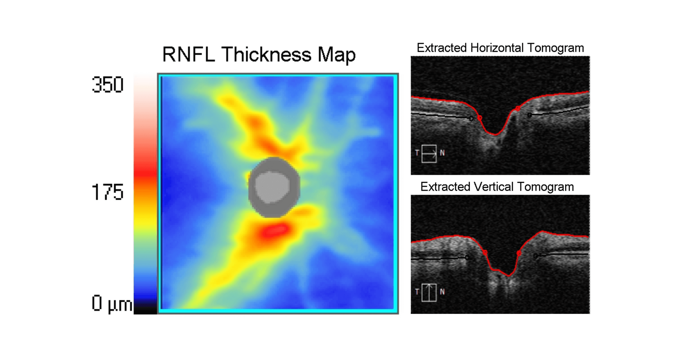
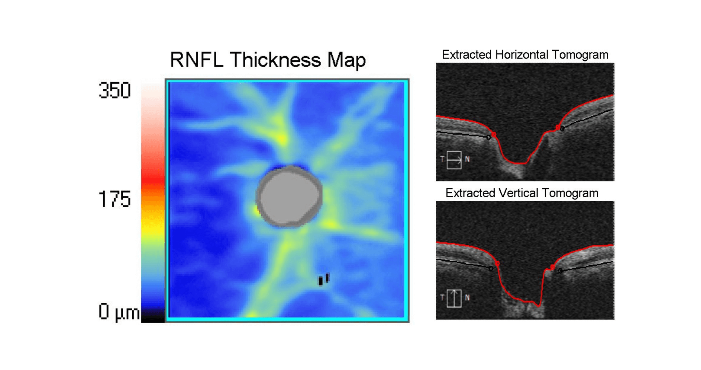
In the early stages of glaucoma, we start to see a thinning of the RNFL which can be seen on the image above. Much of the typical yellow and red colours seen in our “normal” example has gone, being replaced by blue areas representing a thinner RNFL. This indicates that the eye pressure in that eye is now gradually damaging the optic nerve. Our vertical tomogram shows a subtle change in the contour of the cup with the excavation appearing deeper and more “squared” in appearance. The loss of the RNFL causes this change in the shape of the cup and is typical of glaucoma.
We also start to see some functional changes in vision. These changes typically include a reduction in the eye’s sensitivity to light nasally (for example: If looking with the Right eye, the area of visual field around your nose is now unable to detect light unless it is very bright) as can be seen in our results here. These changes are undetectable using patient symptoms alone at this stage unless computerised testing is carried out.
Moderate glaucoma
As glaucoma progresses, the neuroretinal rim becomes thinner.
With moderate glaucoma, we start to see further excavation of the optic cup in the horizontal and vertical tomographs – the dip in the scans look deeper.
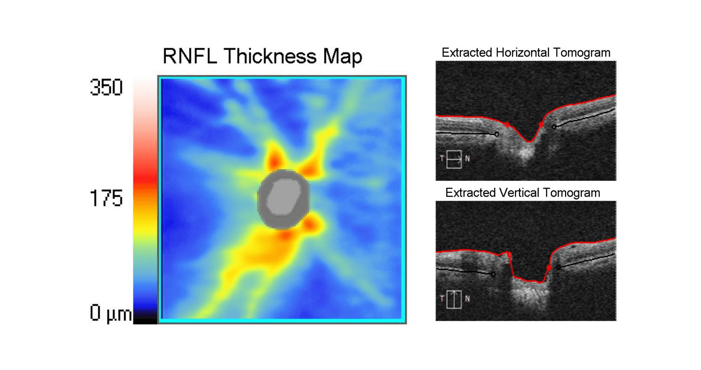
The RNFL thickness map shows further thinning with much of the map now coloured blue. Thinning of the optic nerve is indicative of further advancement of glaucoma and visual field loss.
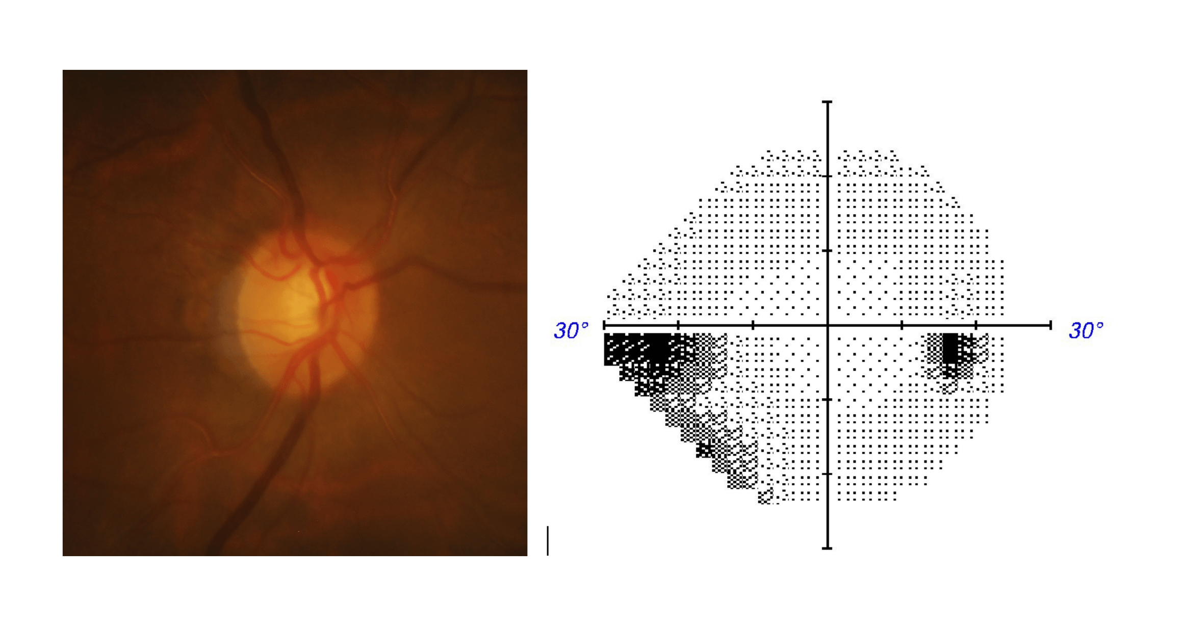
The visual field loss has also extended and deepened (increased area of visual field loss) and is starting to also slightly affect the superior nasal field (top left side of visual field) in this patient. This means that even brighter light is now needed to detect a target on the visual field test. Some areas (such as the black shaded areas) may not detect any light. The area of visual field loss has also expanded in area.
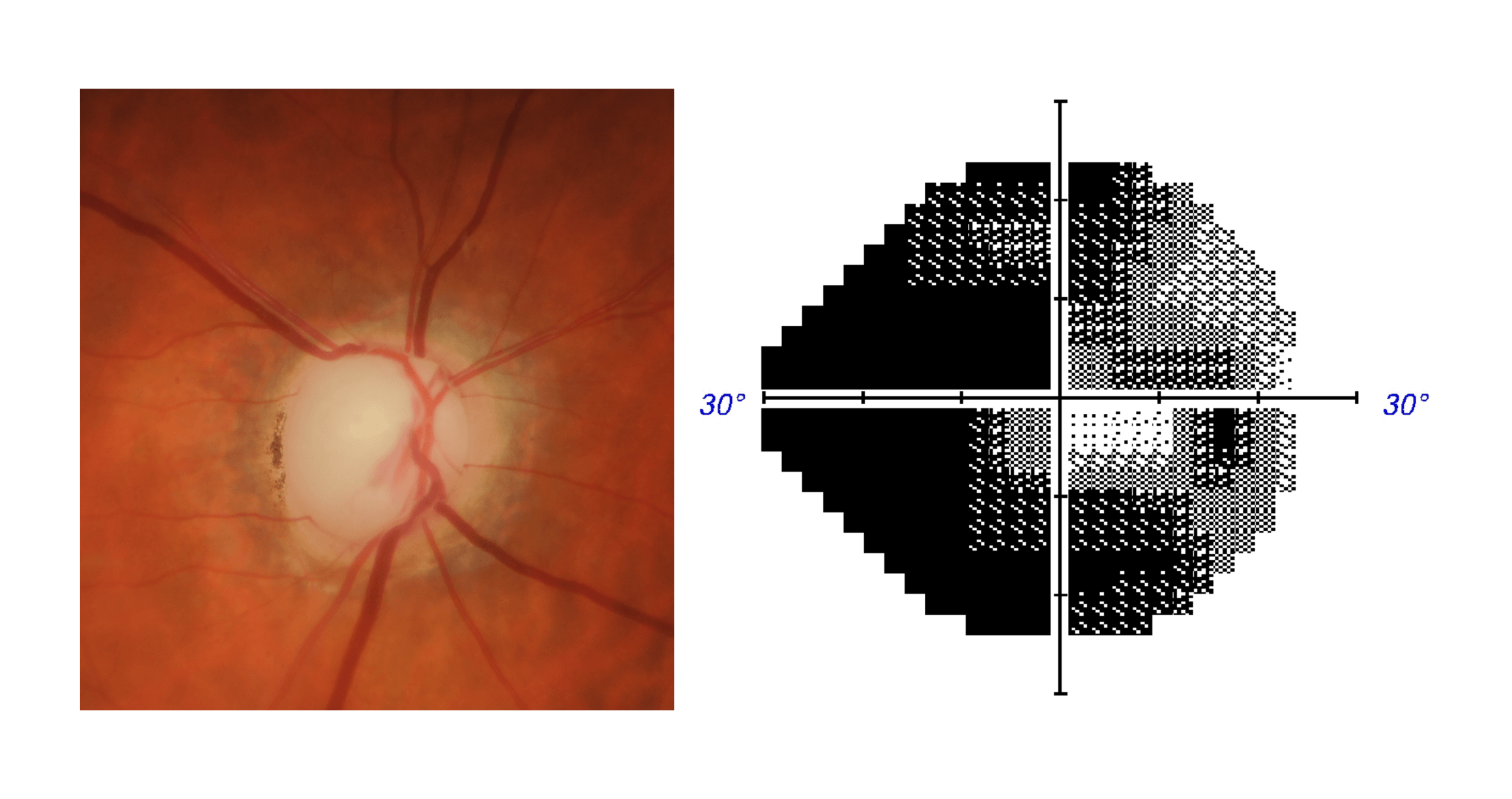
Advanced glaucoma
The optic nerve photo shows almost complete loss of the neuroretinal rim (a dull and pale rim).
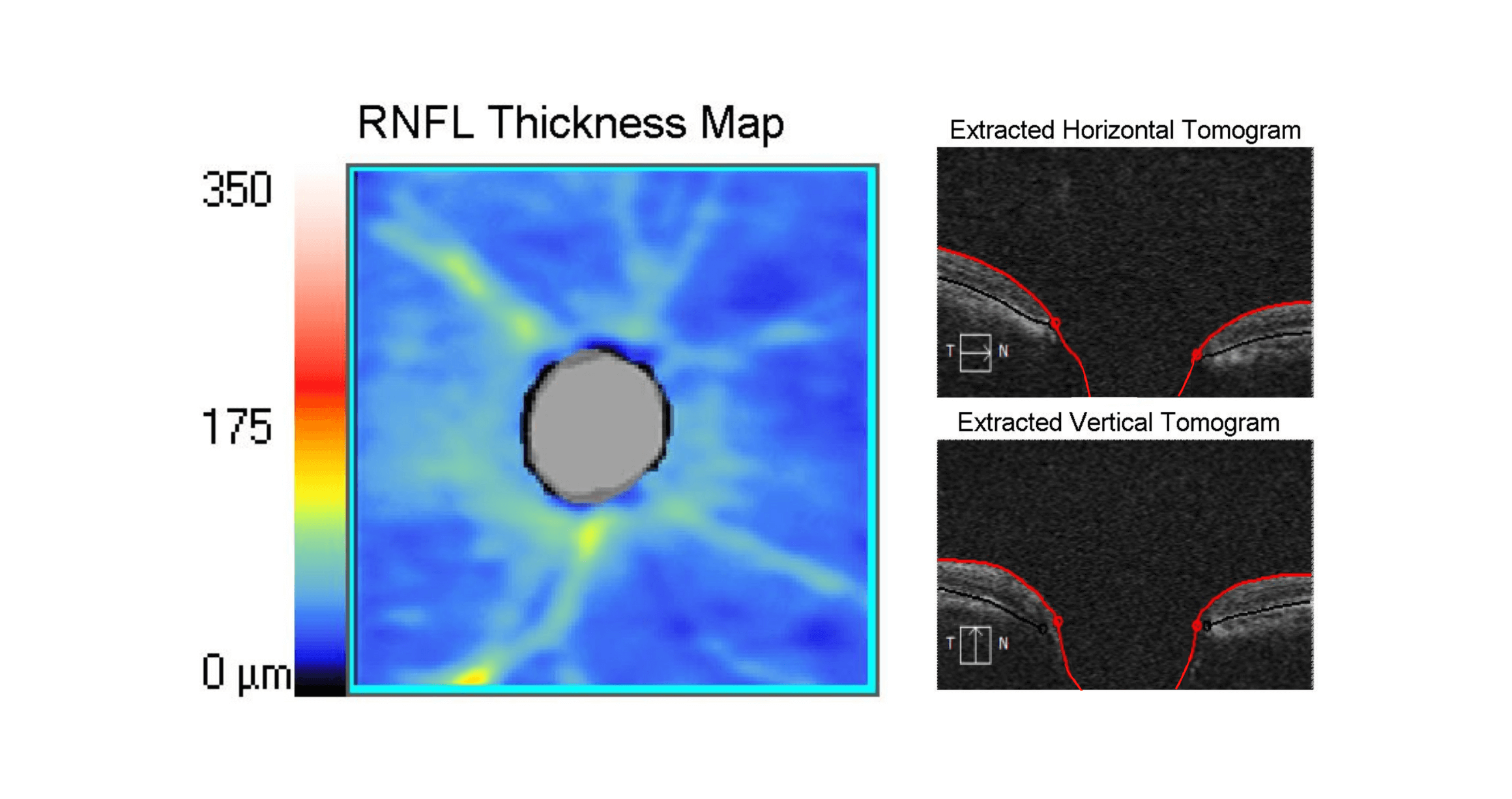
In the advanced stages, there is even further loss of the RNFL as seen (the black and blue indicating very thin or complete loss of RNFL) on the thickness map, and increased excavation of the cup as a result.
Peripheral vision loss becomes more extensive, and the central vision also starts to be affected. At this stage the vision loss can have a significant impact on day to day activities and mobility.
While damage to the optic nerve is irreversible, with early detection and appropriate treatment this extreme vision loss can generally be avoided.
Information and images supplied by The Centre for Eye Health (CFEH)
CFEH is an initiative of Guide Dogs NSW/ACT and the University of New South Wales which provides state-of-the-art diagnostic and disease management services at no cost to patients with the primary goal of reducing preventable blindness in the community.
CFEH has a particular focus on glaucoma and collaborates with a wide range of stakeholders including community optometrists, ophthalmologists and GPs, Prince of Wales and Westmead hospitals and Glaucoma Australia.
CFEH also undertakes cutting edge multi-disciplinary research and educates the optometry profession, working towards reducing preventable blindness.