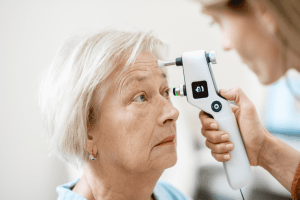
Left untreated glaucoma can cause irreversible vision loss, however early detection and adherence to treatment can halt or significantly slow its progression. The best way to protect your sight is to have a comprehensive eye exam including an optic nerve check which is simple and painless.
During a glaucoma exam your eye health practitioner will:
- Measure your eye pressure, also known as intraocular pressure (IOP).
- Inspect your eye’s drainage angle.
- Examine your optic nerve for damage.
- Test your central and peripheral (side) vision.
- Take a picture or computer measurement of your optic nerve.
- Measure the thickness of your cornea.
It is important to remember that glaucoma cannot be self-detected. Only an optometrist or an ophthalmologist can determine whether you have glaucoma or not.

- Tonometry (Eye Pressure Test)Show lessShow more
Tonometry measures the pressure within the eye. One of the main risk factors for glaucoma is high eye pressure and the best known treatment for glaucoma is lowering the eye pressure. Therefore the accurate measurement of the eye pressure (tonometry) is essential. The Goldman Applanation Tonometer is the most accurate way of measuring the eye pressure. It involves numbing the eye with eye drops first and then the instrument gently contacts the front of the eye to take the measurement.
- Gonioscopy (Drainage Angle Outflow)Show lessShow more
Gonioscopy is the examination of the drainage angle for the outflow of intraocular fluid. Fluid is constantly being made in the eye and it flows out of the eye at the drainage angle. This test can determine if the high eye pressure is caused by a closed/blocked angle (angle closure glaucoma) or if the angle is open but just not working well enough (open angle glaucoma). This is important because the management of each sub-type is slightly different. The test involves putting a mirrored lens on the surface of the eye after using a numbing drop, almost like wearing a contact lens.
- Ophthalmoscopy (Optic Nerve Check)Show lessShow more
Ophthalmoscopy is the visual examination of the optic nerve. Since glaucoma is a disease of the optic nerve, this is a key test. Dilating drops are usually given to enlarge the pupil, so that the optic nerve can be more clearly seen. The nerve(s) will be looked at for signs of glaucoma-related nerve cell loss. This can be done using a table top slit lamp or hand held ophthalmoscope. The appearance of the optic nerve can be documented with a drawing, a photo or with an imaging device. This is so any worsening of the nerve appearance can be detected in the future.
- Perimetry (Visual Field Test)Show lessShow more
Glaucoma initially causes peripheral vision loss that the patient does not notice. This vision loss can be detected with a perimetry test (also known as a visual field test). It involves testing each eye separately with an automated machine that flashes a series of small lights in the periphery to which the patient should react by pressing a button. The perimetry test takes around 3-6 minutes per eye and is repeated 1-2 times a year so that every new test can be compared to the previous ones to look for any worsening.
- Optical Coherence Tomography (OCT)Show lessShow more
An OCT scan is an important test in diagnosing and monitoring glaucoma. It is used to measure the retinal nerve fibre layers (RNFL) around the optic nerve which is an important marker of early to moderate glaucoma damage. For glaucoma suspects and those at the very early stages of glaucoma, the visual field test may not show any peripheral field damage however, the measurements from the OCT can detect early signs of glaucoma. The OCT measurements can also help monitor any changes and progression with glaucoma. This can help determine the treatment plan.
- Pachymetry (Corneal Thickness)Show lessShow more
Pachymetry is a test that measures the thickness of the front window of the eye (cornea). The eye is numbed with drops then a contact probe quickly measures the thickness in a few seconds. A very thick or very thin cornea can affect the pressure/tonometry readings and also a very thin cornea increases the risk of developing glaucoma.
Glaucoma is a complex disease, and no single test can provide enough information to make a diagnosis. The following is a brief overview of the five most common glaucoma tests:
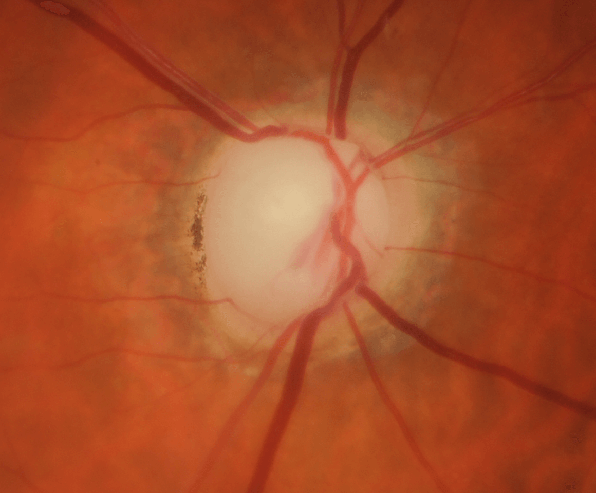
By viewing the images of each stage of glaucoma, we can better understand the progressive loss of the retinal nerve fibre layer (RNFL), and the visual changes associated with this disease.

While anyone may develop glaucoma, some people are at a higher risk including those with a family history of the disease and anyone over the age of 50. Take our quiz to find out if you are at higher risk of developing glaucoma.

'I could not recall if my parents had suffered from glaucoma, but was told to advise my immediate family to have regular eye tests, as there is a high hereditary risk associated with the disease...'